 Search
 Global

ProSound F75 for Obstetrics
ProSound F75 Overview
The ProSound F75 supports various scan methods and a diversified lineup of speciality probes gives you a wide variety of imaging capabilities to meet all your imaging needs.
The new high-efficiency SmartProbes™ are very lightweight and designed to be high in energy conversion to transmit high-quality ultrasound beams without raising the temperature on the probe surface.
 - Click to enlarge image")
 - Click to enlarge image")
 - Click to enlarge image") Click image to enlarge
Click image to enlarge
 - Click to enlarge image") Click image to enlarge
Click image to enlarge
 - Click to enlarge image")
 - Click to enlarge image") Click image to enlarge
Click image to enlarge
 - Click to enlarge image")
 Image Optimizer OFF
 Image Optimizer ON


ProSound F75 Image Technology
Clear images and advanced functions are what you have come to expect from Hitachi – Aloka. Our 60 years of experience and innovation continues with the ProSound F75 platform.
- Compound Pulse Wave Generator (CPWG) – The most advanced broadband beam-forming technology combined with high speed image processing that allows for higher definition ultrasound imaging than ever before.
- Broadband Harmonics™ (BbH) – Provides high quality imaging using an expanded range of harmonic signals. This technology results in excellent image resolution and sensitivity and improved penetration.
- Full Aperture Apodization (FAA) – Processes signals on all channels which significantly enhances focusing and sensitivity and allows for greater image uniformity.
- Adaptive Image Processing (AIP) – Clearly displays differences in tissues, reducing speckle noise while maintaining the frame rate. It can also display outlines more clearly by selectively emphasizing boundaries.
- Spatial Compound Imaging (SCI) – The ultrasound beam is transmitted and received in real time and in the multiple directions resulting in a reduction of speckle noise, suppression of artifacts, and improvement of contrast resolution allowing lesions to be clearly observed.
- Image Optimizer – At the touch of a button the B-mode image is instantly optimized to the user’s preference. This technology continually monitors the user’s typical settings to optimally adjust the image when pressed resulting in less manual adjustments and more efficient examinations.
- SmartProbes – The new high-efficiency probes are very lightweight and designed to be high in energy conversion to transmit high-quality ultrasound beams without raising the temperature on the probe surface.
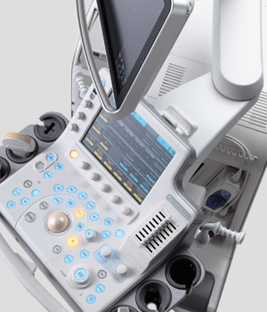
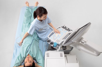
ProSound F75 Ergonomics
3–directional simultaneous adjustment of the control panel
The operation panel of the ProSound F75 can be adjusted so that the switch layout matches the angle of the examiner's arm for comfortable examination. This panel moves sideways and back and forth, and can also be swiveled, fully adjusting to match the examiner's posture.
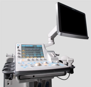
Flexible monitor arm
The angle, height, and distance of the monitor can be optimized for the examiner. The wide-view 19-inch high-resolution monitor is easy to view even from an acute angle.
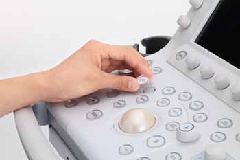
Operation panel is less than 28 inches above the floor
The height of the operation panel a key point for comfortable examinations. That of the ProSound F75 can be lowered down to under 28 inches from the floor.
ProSound F75 Features
Real-time 3D/4D
Real-time 3D allows the acquisition, optimization, manipulation and analysis of structures in 3D and Real-time 3D/4D

Spatio-Temporal Image Correlation (STIC)
A complete 3D volume set of one heart beat can be constructed from the rapid-beating fetal heart, enabling the display in Multi-Planar Reconstruction (MPR) and Multi Slice Imaging (MSI). Greatly contributes to detailed evaluations of the fetal heart by enabling observation from various cross sections.

eFLOW
Displays blood flow with directional information at higher frame rates and spatial resolution compared to conventional methods. Detail and accuracy of blood flow information is greatly increased with reduced blooming of color.

Dual Doppler
Performs Doppler measurements at two points simultaneously, which contributes to the evaluation of fetal arrhythmia.

Extended Field of View (EFV)
EFV (or wide view) provides an extended field-of-view image created from a series of real-time images. As the user moves the transducer across the area of interest a larger image is created that provides clearer spatial relationship information of anatomy and structures. This is especially helpful in assessing structures that are larger than the transducer field of view.

Automatic Volume Measurement (AVM)
Automatic Volume Measurement accurately calculates the 3D volume of a structure. (005_OB - Fetal Bladder - AVM) Multi-slice Imaging (MSI) - Creates multiple Multi-planar reconstruction (MPR) images from a 3D volume data and displays the images simultaneously. The MPR images are displayed parallel to each other and assist in the understanding of the internal structure of the 3D image by acquiring the information of multiple planes in a structure.

Multi-slice Imaging (MSI)
Creates multiple Multi-planar reconstruction (MPR) images from a 3D volume data and displays the images simultaneously. The MPR images are displayed parallel to each other and assist in the understanding of the internal structure of the 3D image by acquiring the information of multiple planes in a structure.

Obstetrics Clinical Images

3rd Trimester Profile

Fetal Heart

Umbilical Cord eFlow

3d Fetal Face

3rd Trimester Profile

OB

3D Fetal Spine

Fetal Heart Multislice

3rd Trimester Fetal Brain Anamoly

Fetal Head

Fetal Bladder

3rd Trimester Fetal Face

OB STIC

3rd Trimester Fetal Brain

4D Fetal Hand

Fetal Head

4D FetalFace&Hand

4D UterineAnamoly

OB STIC

FetalHead

3D Uterus MPR

4D FetalFace

FetalHead

4D FetalHand

FetalHeart

4DShading

FetalHead

UmbilicalCord eFlow

FetalBodyProfile

FetalBrain eFlow

4D FetalFace

FetalHead Anamoly

FetalHeart FAM

LegBones DualScreen

Fetal Face 4D Shading

Fetal Foot

FetalHeart eFlow


Specialty
Transducers
-
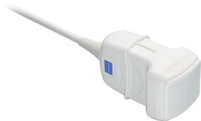
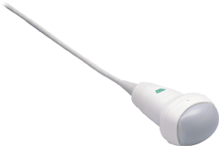 ASU-1010
Convex
10 - 2 MHz
40 mm Radius
60° FOV -
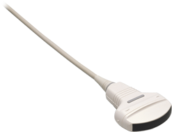
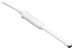
 ASU-1012
Convex
7.5 - 3.75 MHz
10 mm Radius
140° FOV -
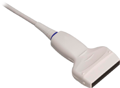
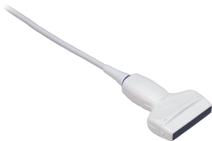
 ASU-1013
Linear
13.33 - 4.44 MHz
40 mm Radius
40° FOV -
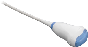 ASU-1014
3D/4D
10 - 2.14 MHz
40 mm Radius
78/60° FOV -
 UST-567
Linear
13.3 - 4.4 MHz
50 mm Width -
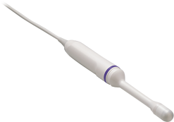
 UST-9118
Convex
7.5 - 3.5 MHz
9 mm Radius
180° FOV -
 UST-9147
Convex
6 - 1.3 MHz
51 mm Radius
71° FOV
Follow Us
For the latest updates and innovations on all of our Women's Health solutions, follow us on:

Contact Info
Sales: 1-800-800-3106
Email: Sales_US@HitachiHealthcare.com