 Search
 Global

ProSound F37 for Gynecology
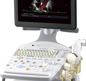
ProSound F37 Overview
Thoroughly simple and compact. As a user-friendly diagnostic ultrasound system full of functional and ergonomic features, F37 is ready to be your partner.
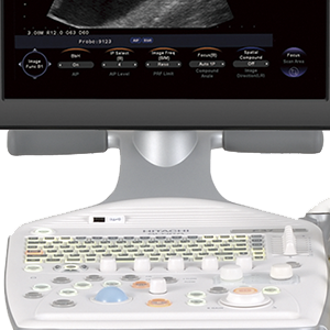
The F37’s simple operational features provide an easy and smooth workflow. The necessary controls for routine examinations are intuitively laid out, with high priority on reducing the examiner's operation steps. Imaging features inherited from higher-class models provide a work environment for concentrated examinations.
 Click image to enlarge
 - Click to enlarge image")
 - Click to enlarge image") Click image to enlarge
Click image to enlarge
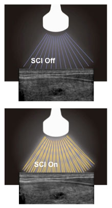
 Image Optimizer OFF
 Image Optimizer ON
ProSound F37 Image Technology
Clear images and advanced functions are what you have come to expect from Hitachi – Aloka. Our 60 years of experience and innovation continues with the ProSound F37 platform.
- Compound Pulse Wave Generator (CPWG) – The most advanced broadband beam-forming technology combined with high speed image processing that allows for higher definition ultrasound imaging than ever before.
- Broadband Harmonics™ (BbH) – Provides high quality imaging using an expanded range of harmonic signals. This technology results in excellent image resolution and sensitivity and improved penetration.
- Adaptive Image Processing (AIP) – Clearly displays differences in tissues, reducing speckle noise while maintaining the frame rate. It can also display outlines more clearly by selectively emphasizing boundaries.
- Spatial Compound Imaging (SCI) – The ultrasound beam is transmitted and received in real time and in the multiple directions resulting in a reduction of speckle noise, suppression of artifacts, and improvement of contrast resolution allowing lesions to be clearly observed.
- Image Optimizer – At the touch of a button the B-mode image is instantly optimized to the user’s preference. This technology continually monitors the user’s typical settings to optimally adjust the image when pressed resulting in less manual adjustments and more efficient examinations.
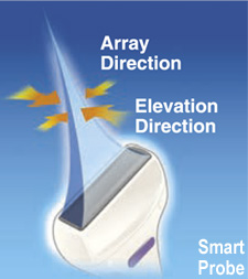
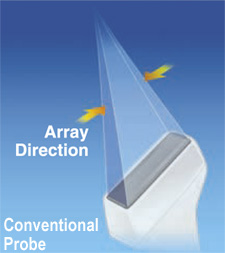
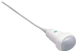
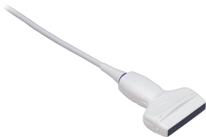
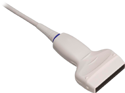
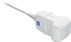
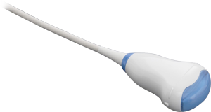
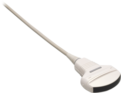
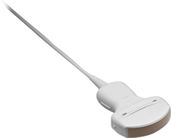
- SmartProbes – The new high-efficiency probes are very lightweight and designed to be high in energy conversion to transmit high-quality ultrasound beams without raising the temperature on the probe surface.

ProSound F37 Ergonomics
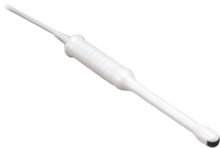
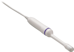
Examinations using the endocavity probe are essential for various gynecological and infertility examinations. With the F37, these examinations have become friendlier to the examiner and patient.
- The image quality contributes to reduction of examination time
- Patient-friendly examinations enabled by the small size of the probe
- Basic measurement results can be transferred onto the report
- Unique probe holder for endocavity probes

ProSound F37 Features
Real-time 3D/4D
Real-time 3D allows the acquisition, optimization, manipulation and analysis of structures in 3D and Real-time 3D/4D

Multi-Planar Reconstruction (MPR)
3D acquisition that provides images of multiple imaging planes including a third plane not accessible with conventional 2D imaging.
")
eFlow
Displays blood flow with directional information at higher frame rates and spatial resolution compared to conventional methods. Detail and accuracy of blood flow information is greatly increased with reduced blooming of color.

Silky Image Processing (SIP)
Reduces artifacts and noise and clarifies tissue borders, enabling easier observation.

Gynecologic Clinical Images

Uterus MPR

Gyn Ovarian Complex Lesion

Gyn Ovarian Cysts

Uterus SIP

4D UterineAnamoly

Uterus

3D Uterus MPR
Build Your Own ProSound F37
Hitachi Healthcare is proud of the reputation we’ve built as an industry leader in Women’s Imaging. Our F37 system is a user-friendly diagnostic ultrasound system full of functional and ergonomic features guaranteed to provide a smooth workflow.
Build your own 2D or 3D system below and find out how much you can save with Hitachi Healthcare. Contact us at 1-800-800-3106 if you have any questions or want to schedule a demonstration.
Build your own F37 ultrasound system, add your accessories, and find out the price in just a few easy clicks. Provide us the following information and we will get started!
Click "Email Quote" for an email copy of your estimate.
Cost does not include applicable state sales tax and shipping charges. Prices valid for U.S. sales only.


Specialty
Transducers
Follow Us
For the latest updates and innovations on all of our Women's Health solutions, follow us on:

Contact Info
Sales: 1-800-800-3106
Email: Sales_US@HitachiHealthcare.com