 Search
 Global

LISENDO 880 for Cardiovascular

LISENDO 880
Hitachi Healthcare pioneered ultrasound for use by cardiologists and we continue to lead the way with the new 2D and 3D premium ultrasound system, the LISENDO 880.
Recognized for our outstanding image quality & Doppler performance, outstanding system reliability and advanced transducer technology, Hitachi Healthcare remains the standard in the field of ultrasound. Hitachi Healthcare’s commitment and dedication to cardiology allows us to offer a wide range of consoles and specifically designed transducers to meet the needs of every cardiologist.
The LISENDO 880 is redefining the vision for cardiovascular ultrasound.
HDAnalytics™
Our advanced HemoDynamic Analytics (HDA) technologies provide a unique and comprehensive evaluation for your patient's cardiovascular health:
- Vector Flow Mapping (VFM) demonstrates flow dynamics in the heart and vessels in a whole new way.
- LV eFLOW is a non-invasive imaging feature that lets you see the endocardial border in the left ventricle with higher sensitivity and resolution than ever before.
- Dual Gate Doppler displays two separate sample gates allowing measurements from two different locations, during the same cardiac cycle.
- 2DTT Provides precise quantification of strain and strain rate to visualize, quantify and analyze regional and global myocardial mechanics using 2D speckle tracking.
- Eyeball EF calculates a Biplane EF from automatic traces on both apical 2 and 4 chamber images.
- eTracking provides multiple parameters necessary for early-stage detection of atherosclerosis.
- Wave Intensity is a new hemodynamic index that provides information about the dynamic behavior of the heart and the vascular system and their interactions.
Premium 3D Imaging
The LISENDO 880 offers Bi-Plane Imaging, 3D Zoom, Active 3D, and Wide Angle 3D live imaging to provide a comprehensive set of data for your 3D and 4D Cardiac Evaluation and Analysis...
Hitachi Healthcare’s cardiology systems provide:
- Extraordinary high-resolution digital imaging with single crystal transducers
- Speckle reduction and edge enhancement technologies providing clearly defined images
- Broadband harmonics offering significantly enhanced sensitivity and axial resolution
- Natural Ergonomics design for reduction of muscle loading while scanning
Cardiovascular Applications
LISENDO 880 Pure Image Technology
The advanced architecture of the LISENDO 880 offers state of the art probe technology for 2D and 3D imaging, a high performance OLED display, premium image optimization parameters such as eFocus and Pure Symphonic Architecture to capture the subtlest of changes and produce exceptional “sound”.
- Advanced Probe Technology – The LISENDO 880 introduces new probe technology to provide improved balance between penetration and resolution. The new S121 offers single crystal probe technology.
- High Performance Display (OLED) – The new OLED display on the LISENDO 880 displays a higher signal to noise ratio and a higher gradient gray scale than with conventional LED displays, providing exceptional resolution.
- Premium Image Optimization Parameters – Parameters like eFocus, NNR, ANR, and HiRez provide detailed optimization control to reduce noise, increase the signal to noise ratio, and improve resolution.
- Pure Symphonic Architecture – We developed high performance image processing engine that provides a variable beamformer and an active backend to deliver state-of-the-art image processing.


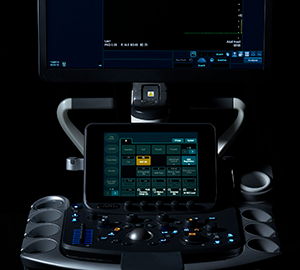
LISENDO 880 Workflow
The LISENDO 880 was designed to provide maximum scanning comfort, along with state-of-the-art technology to help you complete your exams more easily. The systems flexible positioning, including an adjustable panel height and a four point articulating arm, support comfortable operation while the operation panel enables ergonomic function adjustment as a part of our intuitive user interface. Our Smart Cardiac Measurements provide automated analysis to enhance examination efficiency.
The LISENDO 880 delivers seamless workflow users expect in a premium ultrasound system.
The importance of ergonomically designed ultrasound systems cannot be understated. The LISENDO 880 continues our commitment to design systems to minimize repetitive stress while maximizing flexibility across your hospital.
AUTOMATED ANATOMICAL AND STRUCTURAL INTELLIGENT MEASUREMENTS
Auto LV, LA, and RA Volumes and FAC
Left Ventricular, Left Atrium and Right Atrium volumes the Fractional Area Change in the Right Ventricle are automatically measured
Auto LA/Ao
LA diameter in systole and Aortic diameter in Diastole are detected and measured automatically
Auto EF
Automated Teichholz ejection fraction is measured in 2D or M-Mode.
Beat Mode
Automated detection of End Diastolic & End Systolic frames based on the R wave
Protocol Assistant
Move through your study protocol efficiently with automated progressions of modes, measurements, and annotations.
LISENDO 880 Features
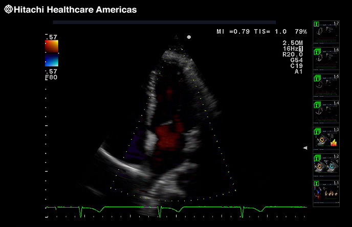
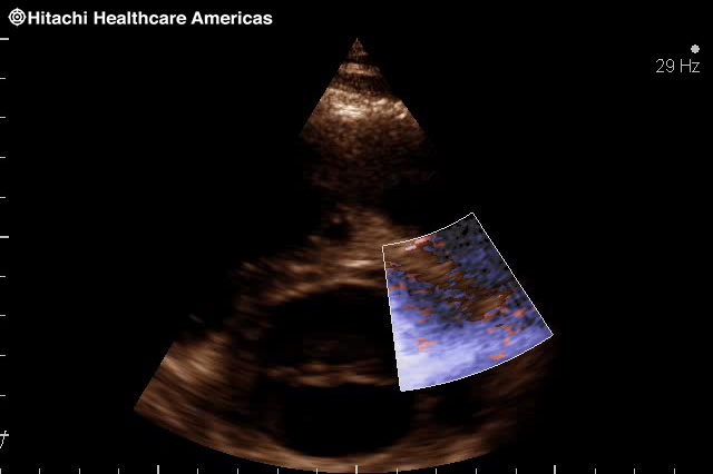
LV eFlow
LV eFLOW is a non-invasive imaging feature that lets you see the endocardial border in the left ventricle with higher sensitivity and resolution than ever before. This high-definition blood flow imaging mode drastically improves spatial and temporal resolution to discriminate blood flow from tissue, enabling endocardial border visualization with one button.
LV eFLOW offers non- directional and directional display. Directional display is useful for identifying endocardial blood flow direction, overcoming the problem of trade-off between high detectability of low velocity flows and aliasing.


click images to enlarge
Dual Gate Doppler
The Dual Gate Doppler generates a full FFT analysis and display from two separate sample gates allowing measurements from two different locations, during the same cardiac cycle. For example:
PW/PW:
- Measure isovolumetric contraction & relaxation time for the evaluation of heart failure patients
PW/TDI:
- E-wave, A-wave and e'- E/e', as a way of evaluating diastolic dysfunction
- Evaluation of atrial fibrillation patients
- R-R Navigation: arrhythmia evaluations
TDI/TDI:
- Useful in evaluation of dyssynchrony and cardiac resynchronization therapy


click images to enlarge
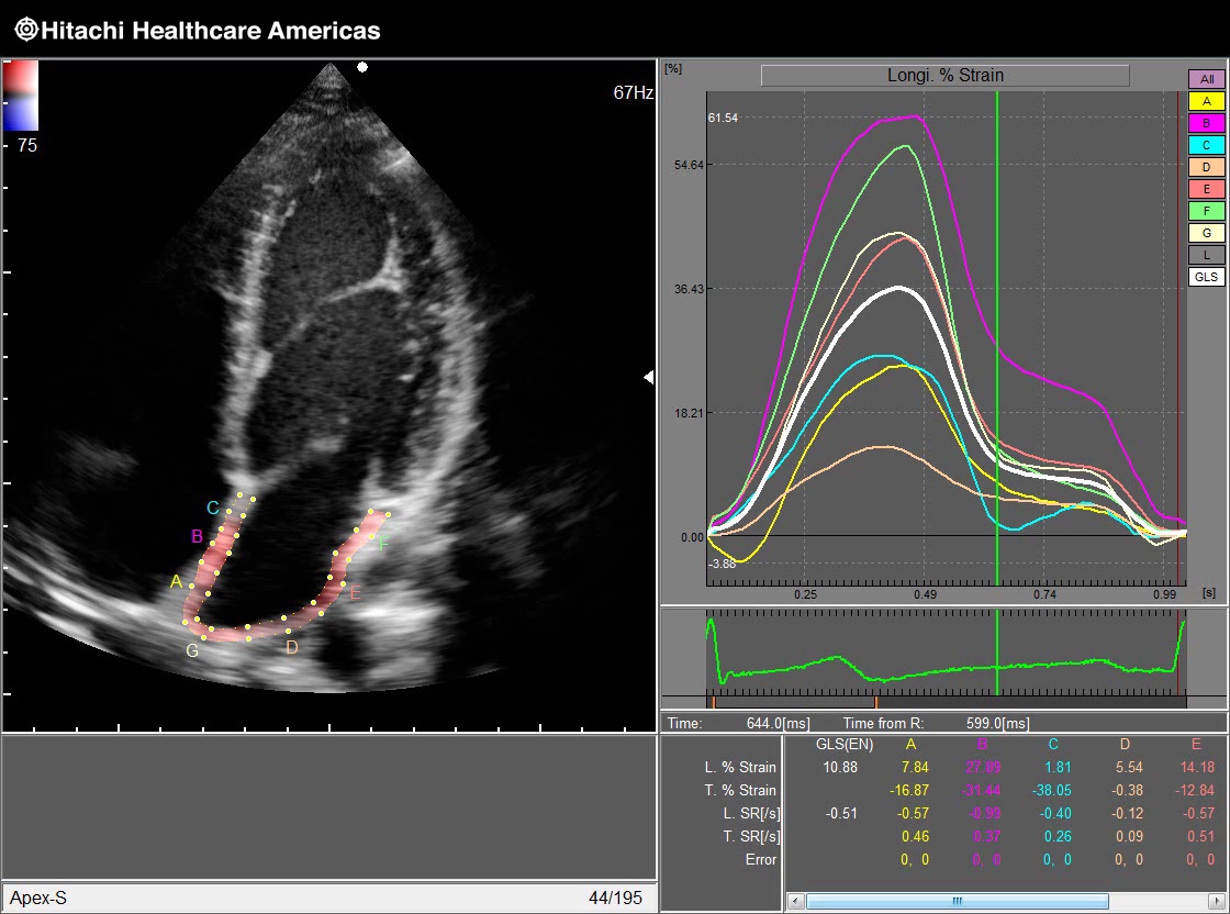
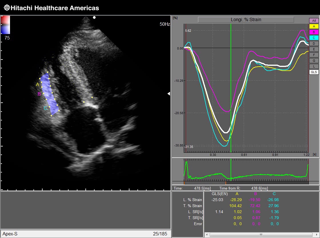
2D Tissue Tracking (2DTT) utilizing speckle technology
Provides precise quantification of strain and strain rate to visualize, quantify and analyze the regional and global myocardial mechanics using 2D speckle tracking.
The 2DTT provides precise quantitative measurements and information such as:
Global Longitudinal Strain/Strain Rate and Radial Strain/Strain Rate Twist angle, displacement, wall thickening and various other parameters to visualize, quantify and analyze the myocardial mechanics.


Strain/Strain Rate
Conventional cardiac function analysis is focused on diagnosis of global heart movement. By opposition, Strain quantifications aim is to analyse cardiac function more locally and sensitively by focusing attention on myocardium areas themselves. Read More >
 Strain |
 Strain Rate |
Free Angular M-mode (FAM)
FAM compares wall motion at multiple locations simultaneously. Three M-mode lines can be set at any position and angle for diagnostic evaluation of wall motion within the same heart cycle. FAM is available in real-time imaging and post imaging from a stored clip.

Stress Echo
With the Hitachi Healthcare Stress Echo you can perform complex Stress Echo examinations smoothly by pressing a single switch to acquire a series of images. The large capacity Cine Memory lets you capture moving images for approximately five continuous minutes in the standard display format.
Dynamic images stored during stress echo examination can be analysed. It is possible to shuffle images and reproduce multiple images simultaneously. The user can also score and make a report while comparing images taken before and after stressing.


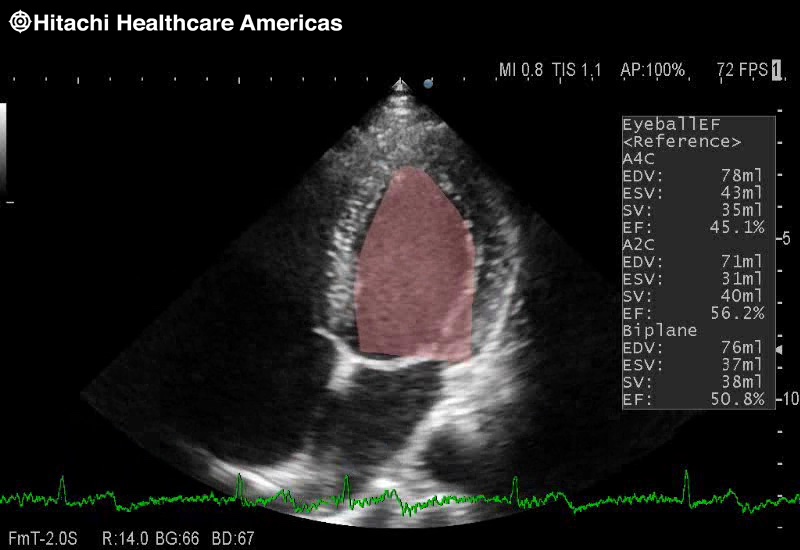
Eyeball EF
Eyeball EF is an automated ejection fraction analysis tool that calculates a Biplane EF from automatic traces of endocardium on both apical 2 and 4 chamber images.
Eyeball EF provides real time Biplane ejection fraction and left ventricular volume based on a built in database of over 1000 multiple tracings.


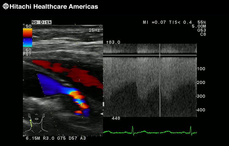
Auto IMT
The relationship between the presence of Carotid atherosclerosis with increased CIMT and coronary arteries disease is now well established. Our Automated IMT Measurement is an easy and simple non invasive tool to quickly assess the evaluation of the cardiovascular risk, at an early stage.
It is possible to automatically extract the max and mean IMT by simply setting the ROI (region of interest) on a long-axis view of the vessel. Equipped with an exclusive report function, multiple measurement values for each part and time phase can be listed, making comparison easier. The displayed items include: max, min, average, SD, points (How many points are used for the result), Width of ROI and Histogram.

Anatomy
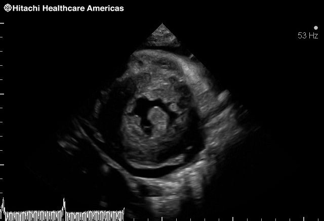
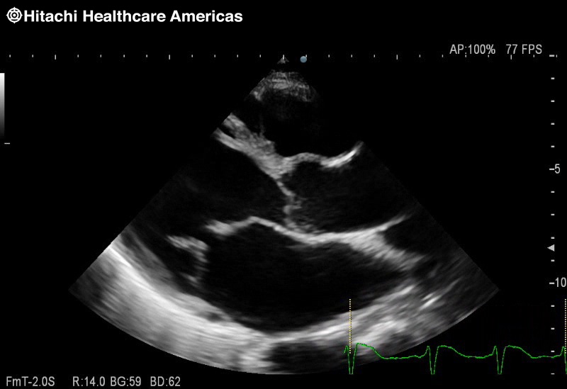
Hypertrophic cardiomyopathy
Hypertrophic cardiomyopathy

Dilated hypertrophic cardiomyopathy
D Shaped septal dampening
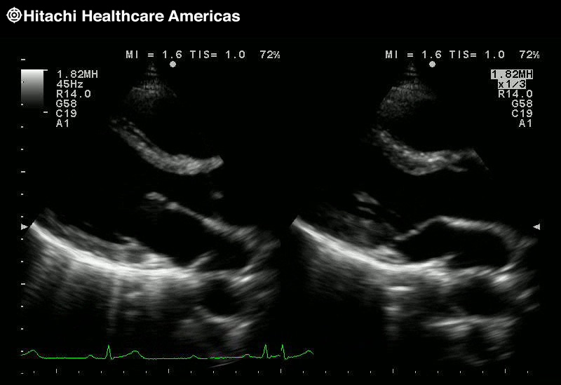
Dual Speed Display

Dual Doppler TDI to TDI

Free Angular M-mode
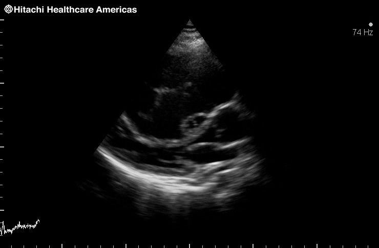
PLAX
Hemodynamics
Color Doppler

Mitral Flow
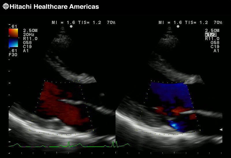
Dual Speed Display Color
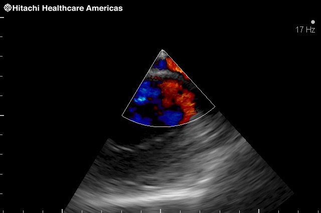
Neonatal Color Doppler

TDI

Dual Doppler TDI to TDI

Dual Doppler PW and TDI

PISA
Coronary eFlow
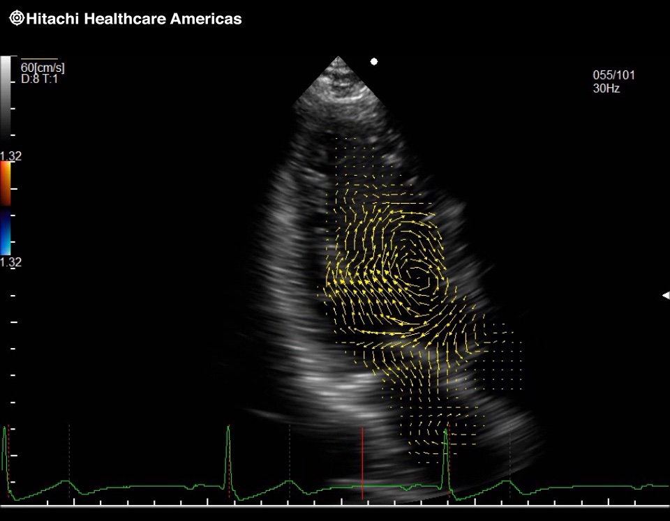
Vector Flow Mapping
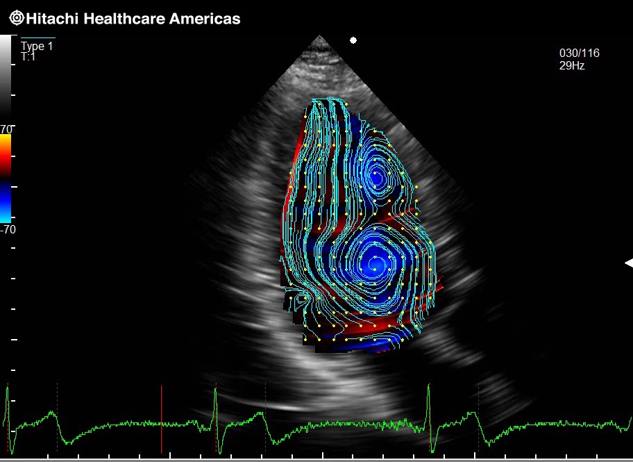
VFM Streamline
eTracking
eTracking Waveforms

Wave Intensity Image

Wave Intensity Graph
Vascular

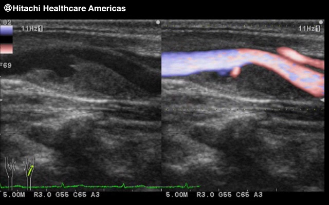
Carotid

Carotid

Carotid Color
Carotid eFlow
CW using linear

DVT

IMT
Wall Motion and Viability

Free Angular M-mode 2 Lines

Free Angular M-mode 3 Lines
EyeBall EF
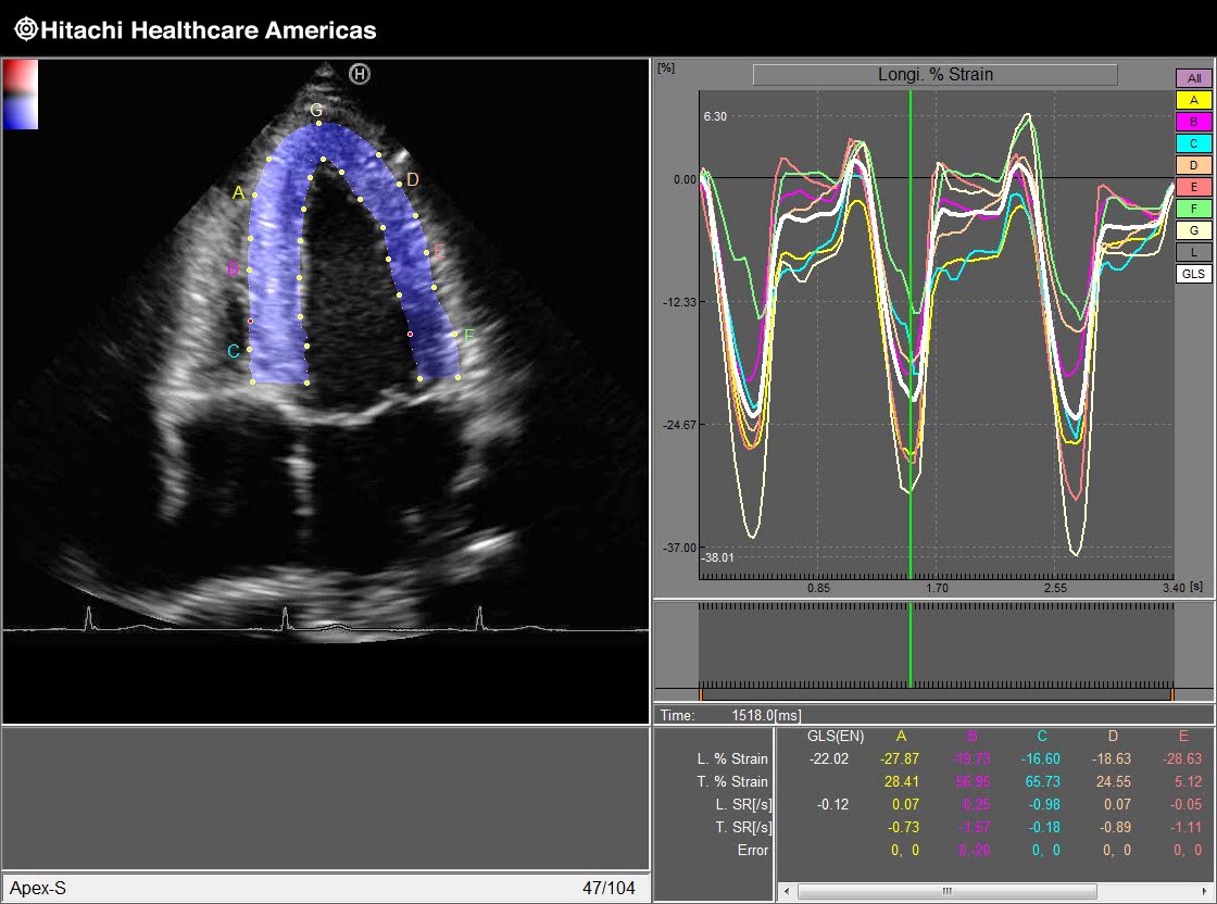
Global Long Strain
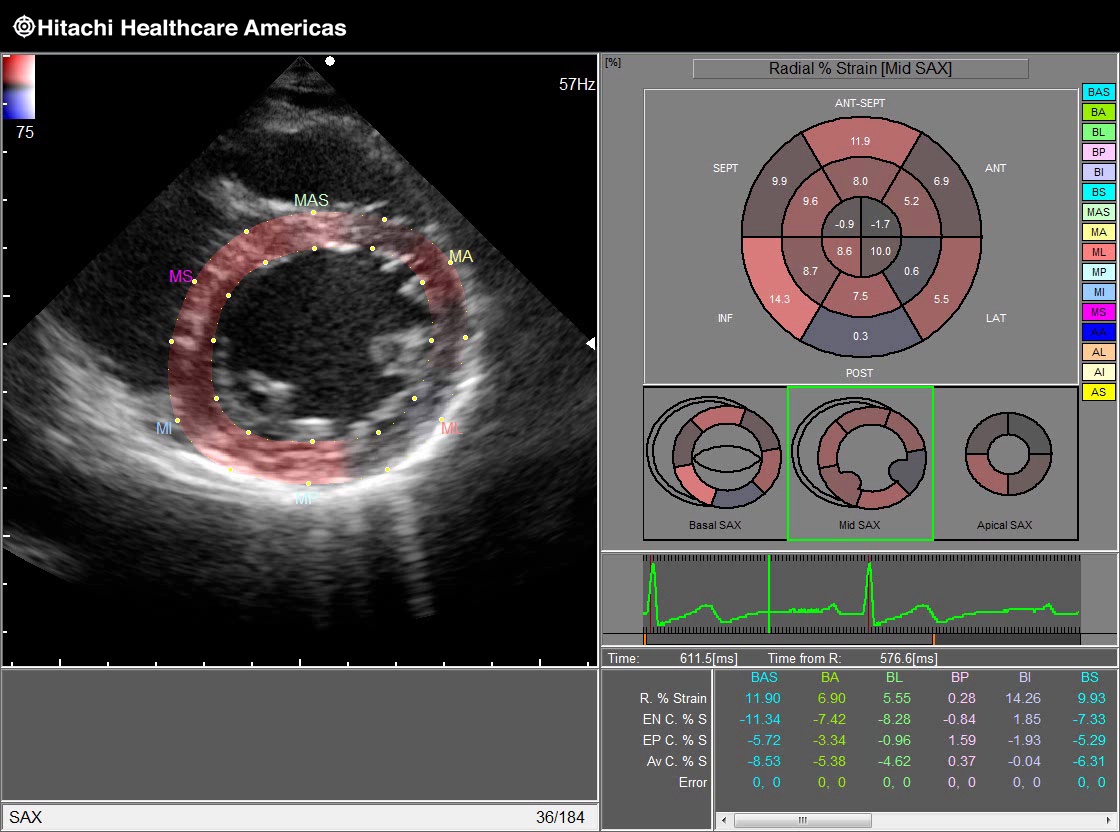
Bull's Eye Mid SAX
LA Strain
RV strain
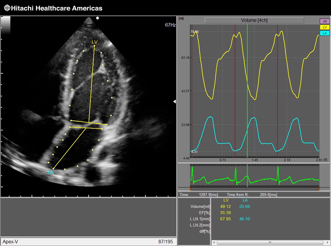
Volume Curves

Stress
LISENDO 880 Advanced Features
Premium 3D Imaging
The LISENDO 880 offers Bi-Plane Imaging, 3D Zoom, Active 3D, and Wide Angle 3D live imaging to provide a comprehensive set of data for your 3D and 4D Cardiac Evaluation and Analysis... Read More >


Vector Flow Mapping (VFM)
Vector Flow Mapping (VFM) evaluates flow dynamics in the heart and vessels in a whole new way. Evaluating pre and post-surgical ventricular and transvalvular blood flow, cardiomyopathies, along with LVAD compliance are just some of the ways VFM may help you access complicated hemodynamics and determine surgical strategies. Using our Arietta 70 ultrasound system and advanced VFM software, new parameters like Vorticity, Energy Loss, Circulation and Shear Wall Stress can be analyzed.


eTRACKING
Comprehensive analysis package supporting the assessment of Atherosclerosis
The severity of Cardiovascular disease is likely to be reduced if atherosclerosis is detected early and before morphologic changes such as plaque and wall thickening are visible in the arterial walls. Prevention and treatment of lifestyle-related diseases are increasingly important today, and so the role of the ultrasound diagnostic system is not only to observe the morphologic changes but also to perform functional assessment... Read More >
![]()
![]()
Wave Intensity
The new Index of Cardiovascular Circulation Dynamics
The heart and the arterial system constantly interact with each other through forward travelling waves and reflected waves. Wave Intensity (WI) is calculated as the product of the derivatives of the simultaneously recorded blood-pressure changes and blood-flow-velocity changes. Wave Intensity can be obtained at an arbitrary point in the circulatory system. Read More >


Specialty
Transducers
-
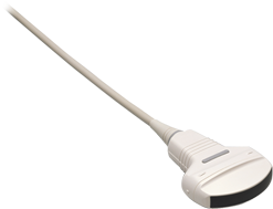
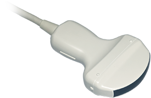
 C251
C251
5 - 1 MHz
50 mm Radius
75° FOV -
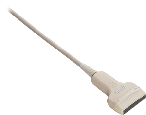
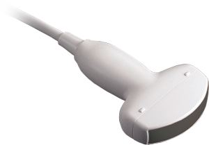
 C35
C35
8 - 2 MHz
70° FOV -
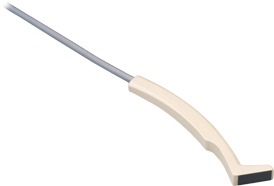
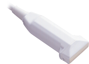 L34
L34
Linear
7 - 3 MHz
38 mm Width -
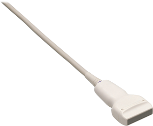
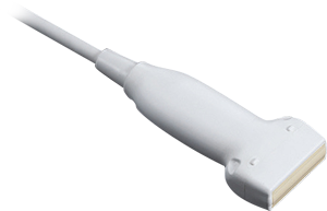 L441
L441
12 - 2 MHz
38 mm -
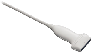 L64
L64
Linear
18 - 5 MHz
38 mm Width -
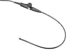
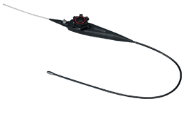
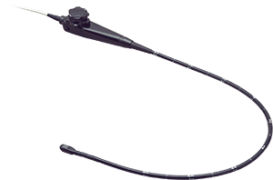 MXS2ESLL
10 - 1 MHz
90° FOV -
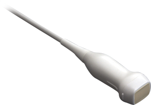
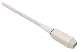
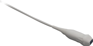
 S121
S121
Sector
5 - 1 MHz
90° FOV -
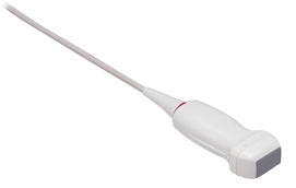
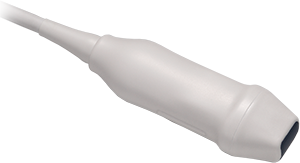
 S31
S31
Sector
9 - 2 MHz
90° FOV -
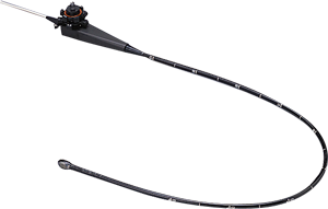
 S3ESEL
7.5 - 3 MHz
90° FOV -
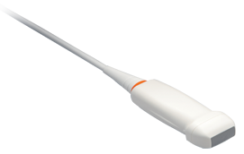
 S42
S42
14 - 3 MHz
90° FOV -
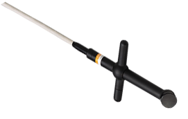 UST-2265-2
Independent CW
Doppler
2 MHz -
 UST-2266-5
Cardiac CW Doppler
5 MHz
Follow Us
For the latest updates and innovations on all of our Cardiovascular solutions, follow us on:


Contact Info
Sales: 1-800-800-3106
Email: Sales_US@HitachiHealthcare.com