 Search
 Global

ARIETTA 65 for Cardiovascular

ARIETTA 65 Overview
Hitachi Healthcare pioneered ultrasound for use by cardiologists and we continue to lead the way with major innovations. Recognized for our outstanding image quality & Doppler performance, outstanding system reliability and advanced transducer technology, Hitachi Healthcare remains the standard in the field of ultrasound.
Hitachi Healthcare's commitment and dedication to cardiology allows us to offer a wide range of consoles and specifically designed transducers to meet the needs of every cardiologist.
HDAnalytics™
Our advanced HemoDynamic Analytics (HDA) technologies provide a unique and comprehensive evaluation for your patient's cardiovascular health:
- LV eFLOW is a non-invasive imaging feature that lets you see the endocardial border in the left ventricle with higher sensitivity and resolution than ever before.
- 2DTT Provides precise quantification of strain and strain rate to visualize, quantify and analyze regional and global myocardial mechanics using 2D speckle tracking.
- eTracking provides multiple parameters necessary for early-stage detection of atherosclerosis.
- Wave Intensity is a new hemodynamic index that provides information about the dynamic behavior of the heart and the vascular system and their interactions.
Hitachi Healthcare's cardiology systems provide:
- Extraordinary high-resolution digital imaging with single crystal transducers
- Speckle reduction and edge enhancement technologies providing clearly defined images
- User defined and customizable study protocols guaranteeing exam consistency
Cardiovascular Applications
") Click image to enlarge
Click image to enlarge
 AIP OFF
 AIP ON
 SCI OFF
 SCI ON
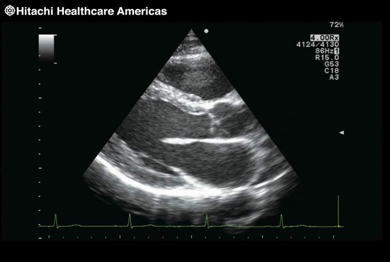 Image Optimizer OFF
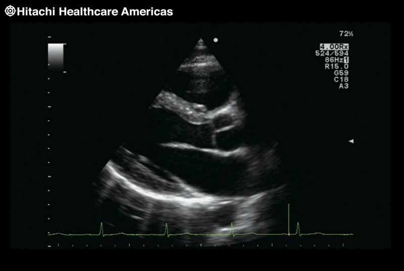 Image Optimizer ON


ARIETTA 65 Image Technology
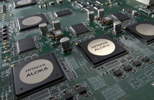
Clear images and advanced functions are what you have come to expect from Hitachi Aloka. Our 60 years of experience and innovation continues with the ARIETTA 65 platform.
- Compound Pulse Wave Generator Plus (CPWG+) – The most advanced broadband beam-forming technology combined with high speed image processing that allows for higher definition ultrasound imaging than ever before.
- Symphonic Technology – Provides high quality imaging using an expanded range of harmonic signals. This technology results in excellent image resolution and sensitivity and improved penetration.
- HI REZ – Clearly displays differences in tissues, reducing speckle noise while maintaining the frame rate. It can also display outlines more clearly by selectively emphasizing boundaries.
- Compound Imaging – The ultrasound beam is transmitted and received in real time and in the multiple directions resulting in a reduction of speckle noise, suppression of artifacts, and improvement of contrast resolution allowing lesions to be clearly observed.
- Image Optimizer – At the touch of a button the B-mode image is instantly optimized to the user’s preference. This technology continually monitors the user’s typical settings to optimally adjust the image when pressed resulting in less manual adjustments and more efficient examinations.
- Single Crystal Probes – A single crystal is used to provide the piezoelectric elements of the probe. Single crystal technology achieves higher sensitivity and wider bandwidths over standard piezoceramics.

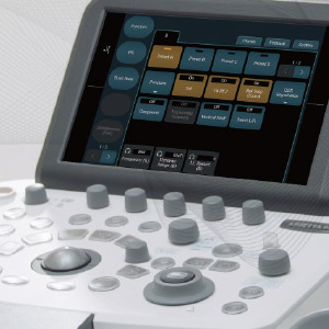
ARIETTA 65 Ergonomics
ARIETTA 65 is ergonomically designed to allow the examiner to scan in comfort irrespective of the type of patient or clinical examination. The adjustment of the panel height between 70 and 100 cm is one of the key contributory elements.
IPS-Pro (In-Plane Switching) LCD panel technology
ARIETTA 65 is fitted with IPS-Pro monitor, giving a high quality display of the images from a wide viewing angle.
Lighter by 25%
ARIETTA 65, almost 25% lighter in weight than conventional systems (Hitachi Healthcare in-house comparison), can be moved around with little effort and operated easily in confined spaces.
User-friendly operation panel
The console layout is arranged to provide intuitively smooth operation, with a large palm rest provided centrally to give optimum wrist support.


ARIETTA 65 Features
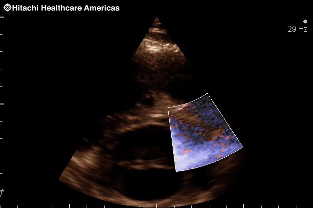
LV eFlow
LV eFLOW is a non-invasive imaging feature that lets you see the endocardial border in the left ventricle with higher sensitivity and resolution than ever before. This high-definition blood flow imaging mode drastically improves spatial and temporal resolution to discriminate blood flow from tissue, enabling endocardial border visualization with one button.
LV eFLOW offers non- directional and directional display. Directional display is useful for identifying endocardial blood flow direction, overcoming the problem of trade-off between high detectability of low velocity flows and aliasing.


click images to enlarge
Dual Gate Doppler
The Dual Gate Doppler generates a full FFT analysis and display from two separate sample gates allowing measurements from two different locations, during the same cardiac cycle. For example:
PW/PW:
- Measure isovolumetric contraction & relaxation time for the evaluation of heart failure patients
PW/TDI:
- E-wave, A-wave and e'- E/e', as a way of evaluating diastolic dysfunction
- Evaluation of atrial fibrillation patients
- R-R Navigation: arrhythmia evaluations
TDI/TDI:
- Useful in evaluation of dyssynchrony and cardiac resynchronization therapy


click images to enlarge
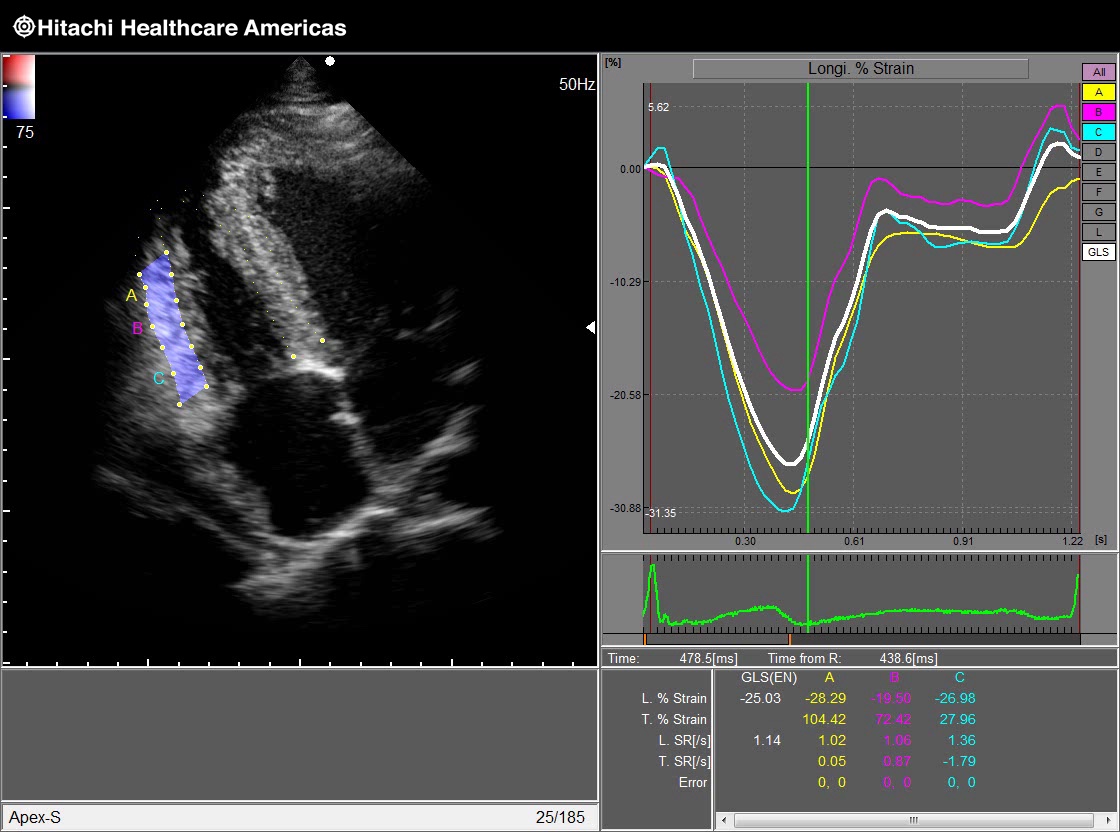
2D Tissue Tracking (2DTT) utilizing speckle technology
Provides precise quantification of strain and strain rate to visualize, quantify and analyze the regional and global myocardial mechanics using 2D speckle tracking.
The 2DTT provides precise quantitative measurements and information such as:
Global Longitudinal Strain/Strain Rate and Radial Strain/Strain Rate Twist angle, displacement, wall thickening and various other parameters to visualize, quantify and analyze the myocardial mechanics.


Strain/Strain Rate
Conventional cardiac function analysis is focused on diagnosis of global heart movement. By opposition, Strain quantifications aim is to analyse cardiac function more locally and sensitively by focusing attention on myocardium areas themselves. Read More >
 Strain |
 Strain Rate |
Free Angular M-mode (FAM)
FAM compares wall motion at multiple locations simultaneously. Three M-mode lines can be set at any position and angle for diagnostic evaluation of wall motion within the same heart cycle. FAM is available in real-time imaging and post imaging from a stored clip.

Stress Echo
With the Hitachi Healthcare Stress Echo you can perform complex Stress Echo examinations smoothly by pressing a single switch to acquire a series of images. The large capacity Cine Memory lets you capture moving images for approximately five continuous minutes in the standard display format.
Dynamic images stored during stress echo examination can be analysed. It is possible to shuffle images and reproduce multiple images simultaneously. The user can also score and make a report while comparing images taken before and after stressing.


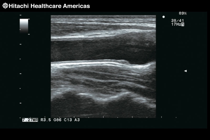
Auto IMT
The relationship between the presence of Carotid atherosclerosis with increased CIMT and coronary arteries disease is now well established. Our Automated IMT Measurement is an easy and simple non invasive tool to quickly assess the evaluation of the cardiovascular risk, at an early stage.
It is possible to automatically extract the max and mean IMT by simply setting the ROI (region of interest) on a long-axis view of the vessel. Equipped with an exclusive report function, multiple measurement values for each part and time phase can be listed, making comparison easier. The displayed items include: max, min, average, SD, points (How many points are used for the result), Width of ROI and Histogram.

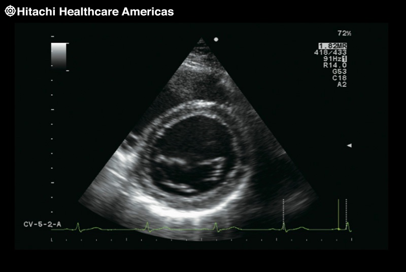
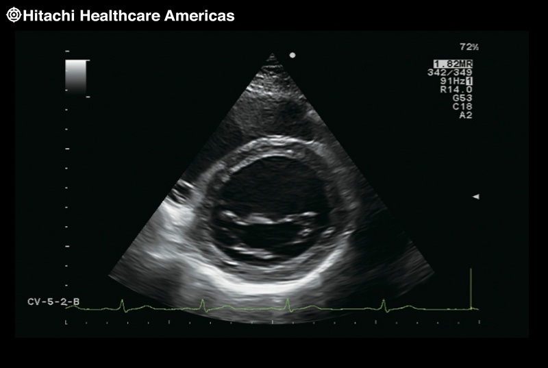
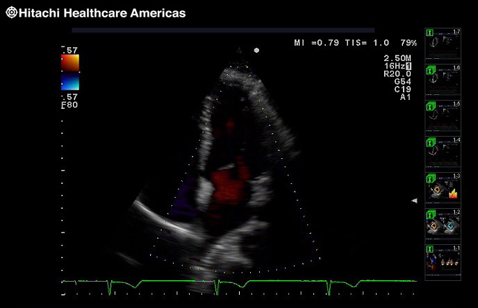
Anatomy
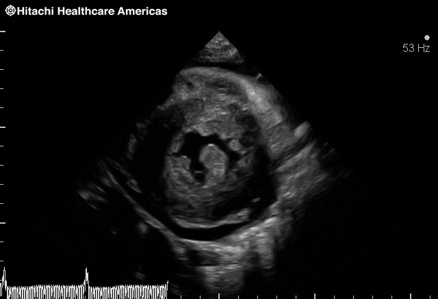
Hypertrophic cardiomyopathy
Hypertrophic cardiomyopathy

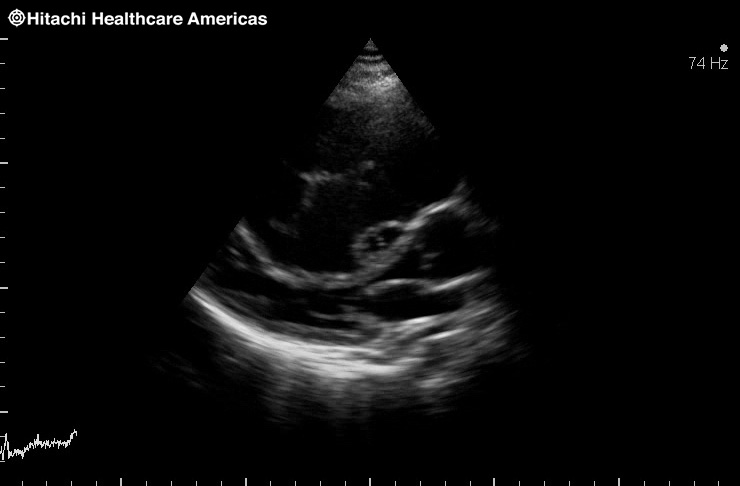
Dilated hypertrophic cardiomyopathy
D Shaped septal dampening
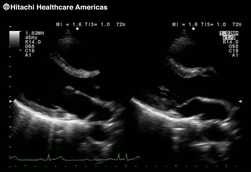
Dual Speed Display

Dual Doppler TDI to TDI

Free Angular M-mode
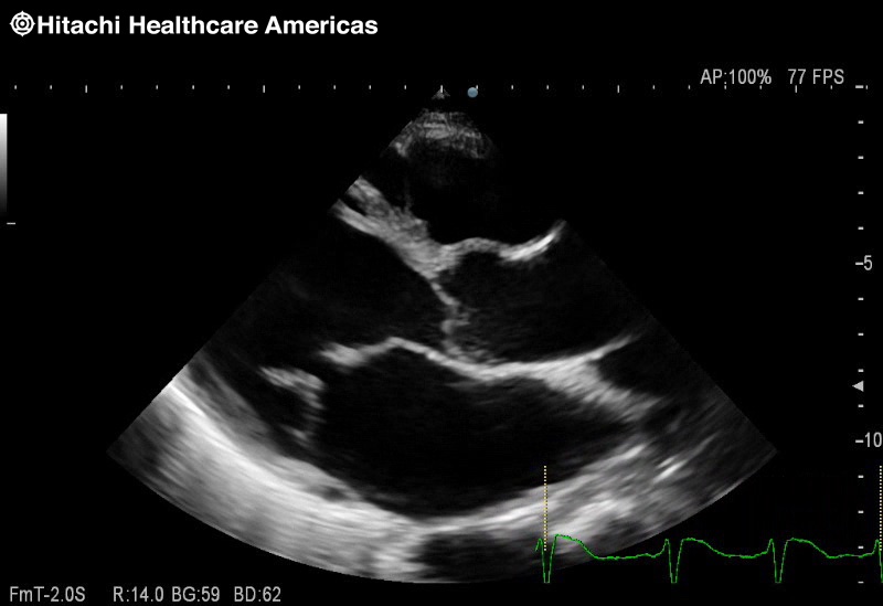
PLAX
Hemodynamics
Color Doppler

Mitral Flow
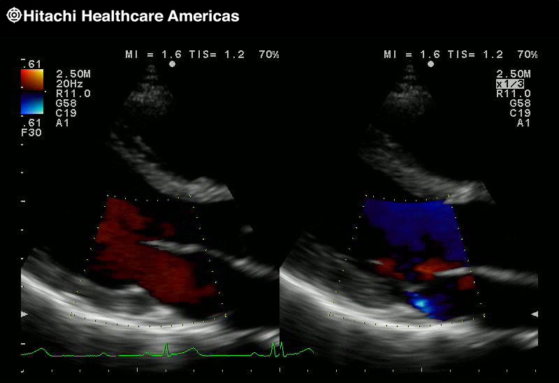
Dual Speed Display Color
Neonatal Color Doppler

TDI

Dual Doppler TDI to TDI

Dual Doppler PW and TDI

PISA
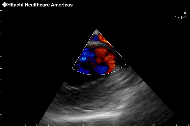
Coronary eFlow
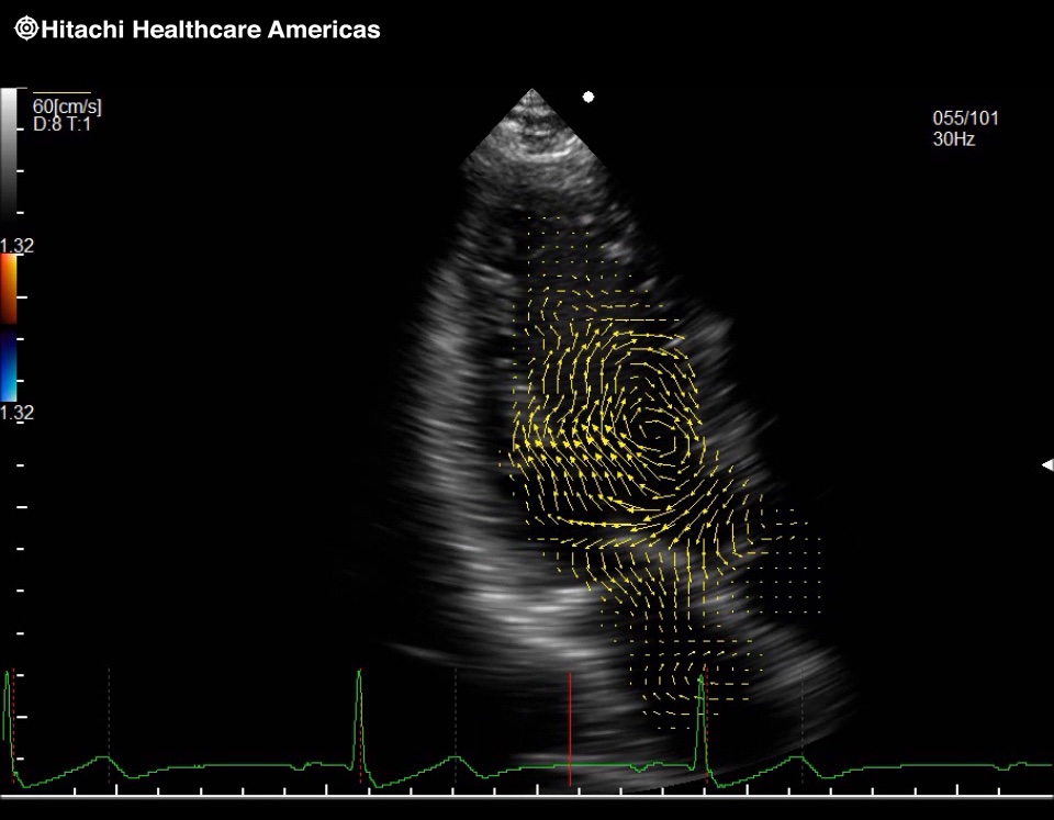
Vector Flow Mapping
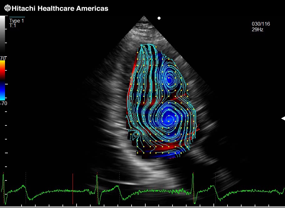
VFM Streamline
eTracking
eTracking Waveforms

Wave Intensity Image

Wave Intensity Graph
Vascular

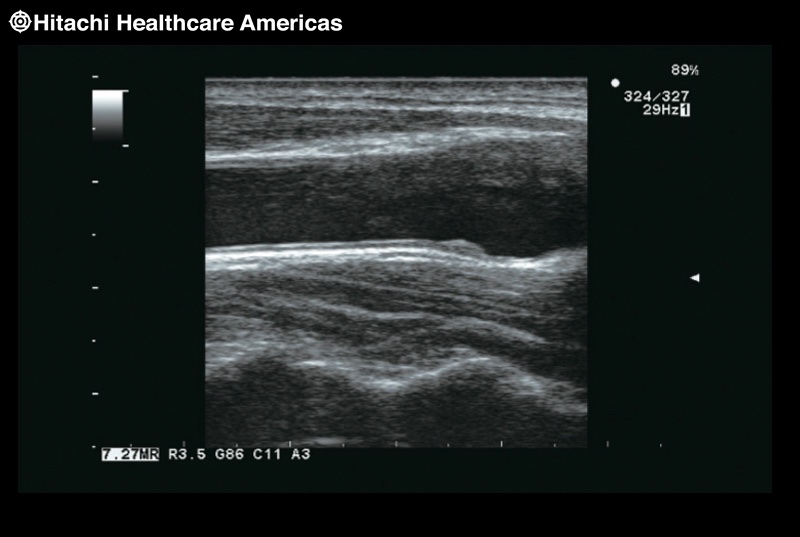
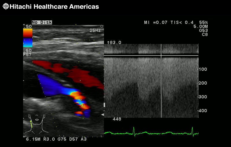
Carotid

Carotid

Carotid Color
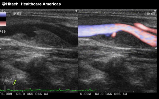
Carotid eFlow
CW using linear

DVT

IMT
Wall Motion and Viability

Free Angular M-mode 2 Lines

Free Angular M-mode 3 Lines
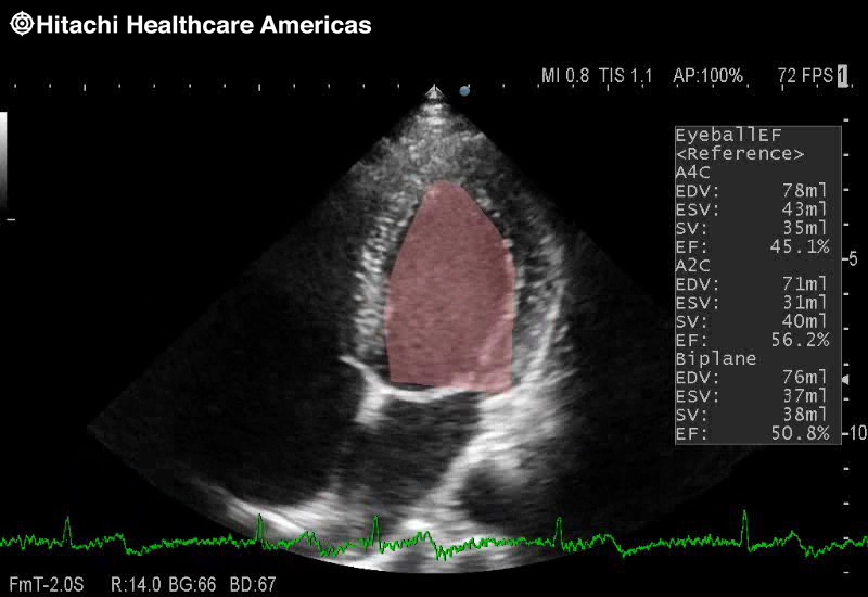
EyeBall EF
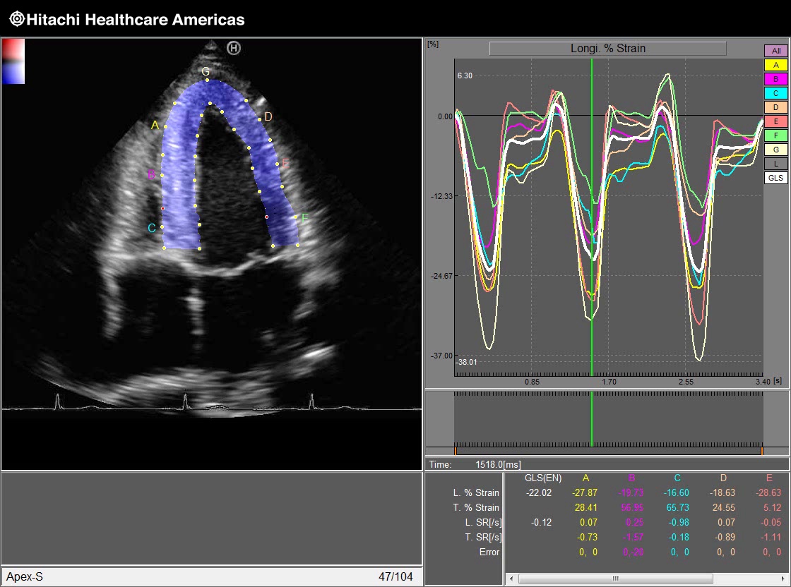
Global Long Strain
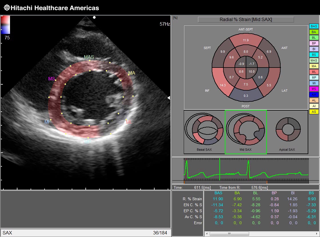
Bull's Eye Mid SAX
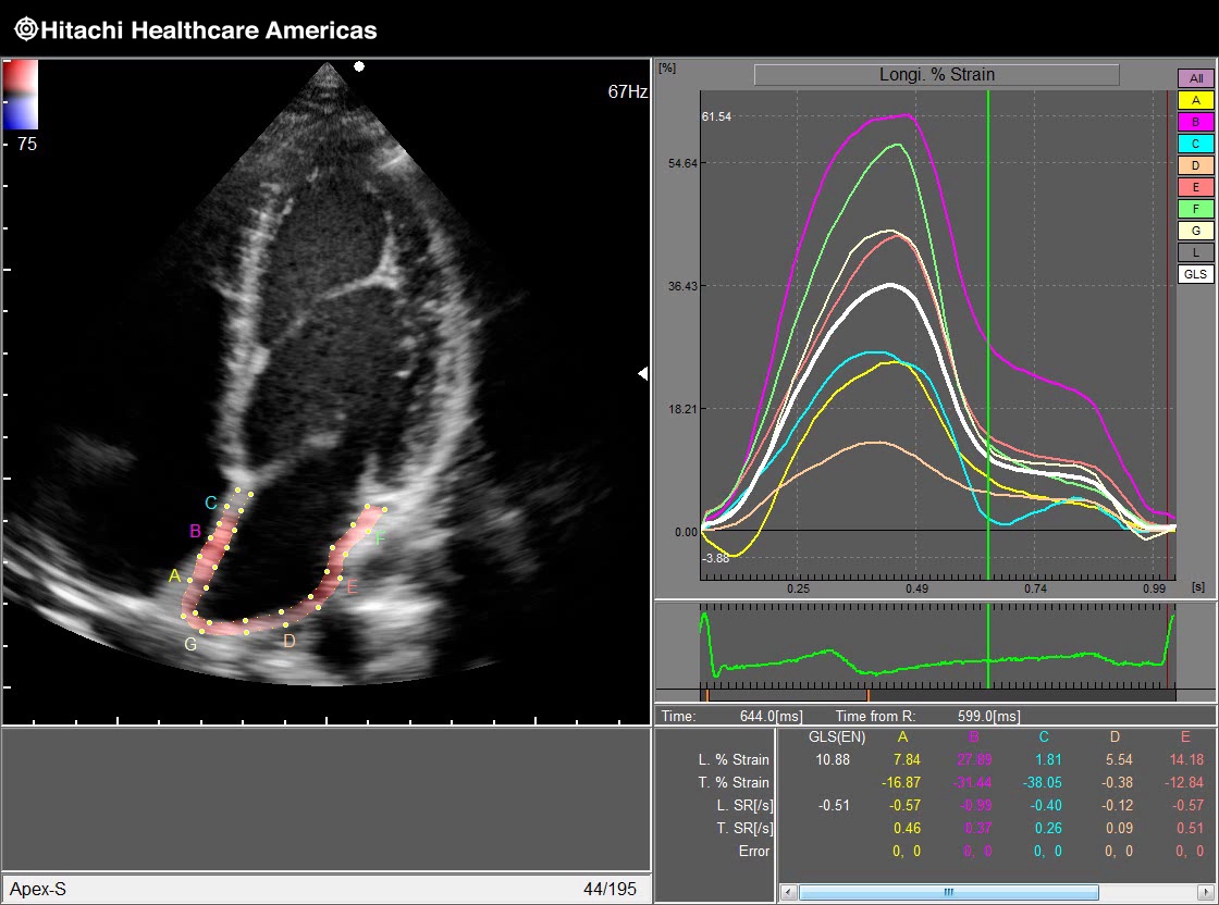
LA Strain
RV strain
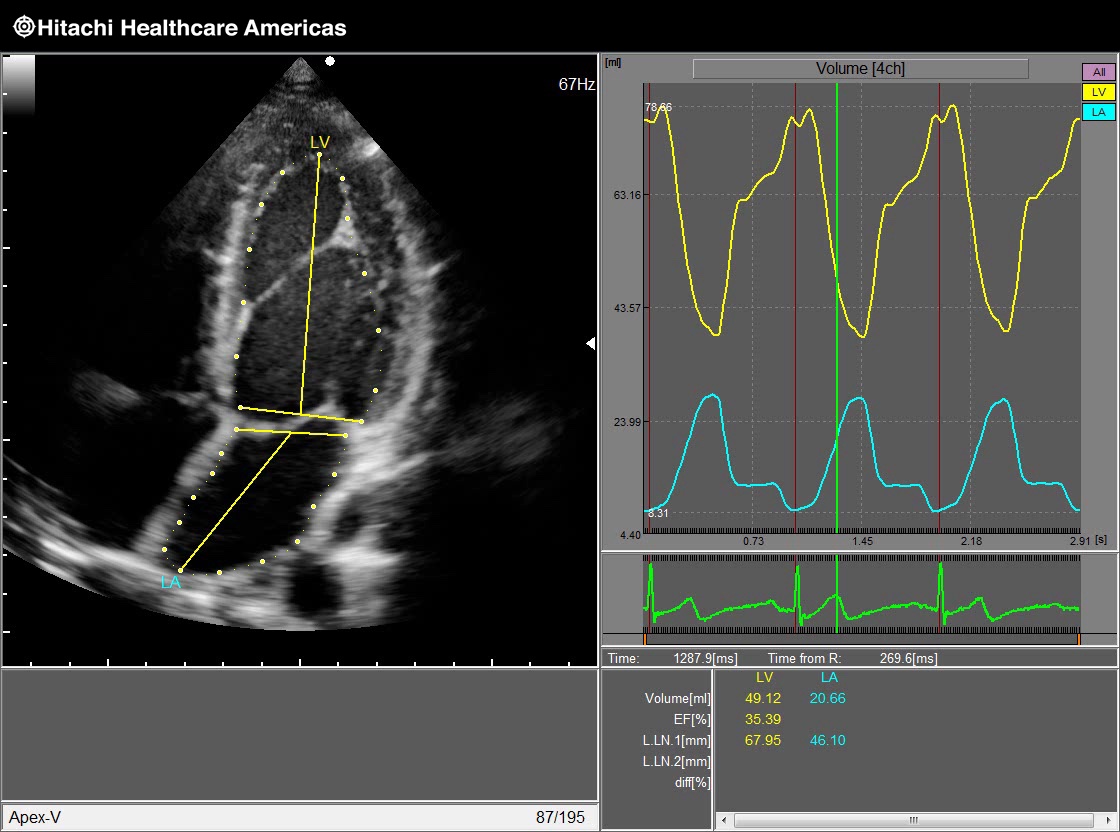
Volume Curves

Stress
ARIETTA 65 Advanced Features
eTRACKING
Comprehensive analysis package supporting the assessment of Atherosclerosis
The severity of Cardiovascular disease is likely to be reduced if atherosclerosis is detected early and before morphologic changes such as plaque and wall thickening are visible in the arterial walls. Prevention and treatment of lifestyle-related diseases are increasingly important today, and so the role of the ultrasound diagnostic system is not only to observe the morphologic changes but also to perform functional assessment... Read More >
![]()
![]()
Wave Intensity
The new Index of Cardiovascular Circulation Dynamics
The heart and the arterial system constantly interact with each other through forward travelling waves and reflected waves. Wave Intensity (WI) is calculated as the product of the derivatives of the simultaneously recorded blood-pressure changes and blood-flow-velocity changes. Wave Intensity can be obtained at an arbitrary point in the circulatory system. Read More >


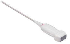
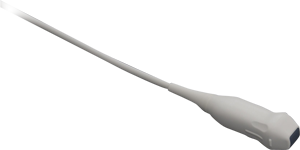
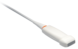
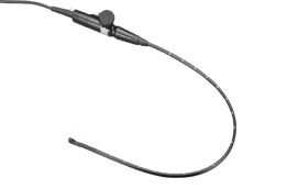

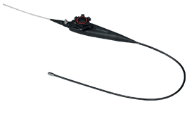

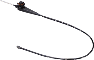
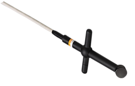
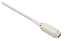
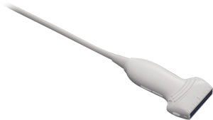
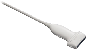
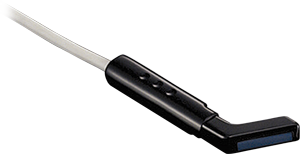
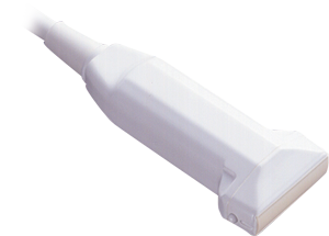
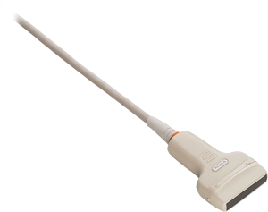
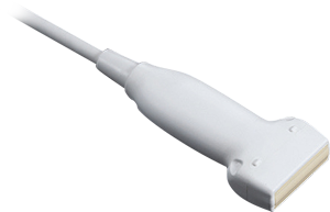
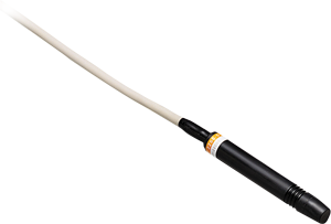
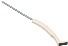
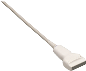
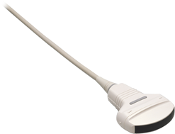
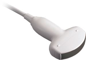
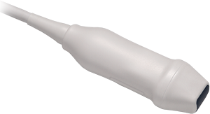
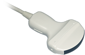
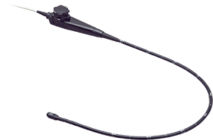

Specialty
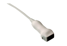
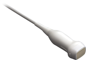
Transducers
Follow Us
For the latest updates and innovations on all of our Cardiovascular solutions, follow us on:

Contact Info
Sales: 1-800-800-3106
Email: Sales_US@HitachiHealthcare.com